Doctor Banner

Oxygen deprivation, or intrapartum asphyxia, can cause Cerebral Palsy. One of the most common types of brain damage caused by oxygen loss is called hypoxic-ischemic encephalopathy, or HIE. When HIE occurs, it often leads to severe developmental or cognitive delays, or motor impairments that become more apparent as the child continues to develop.
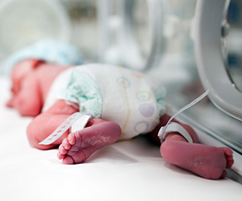
Hypoxic-ischemic encephalopathy, or HIE, is the brain injury caused by oxygen deprivation to the brain, also commonly known as intrapartum asphyxia. The newborn’s body can compensate for brief periods of depleted oxygen, but if the asphyxia lasts too long, brain tissue is destroyed. Hypoxic-ischemic encephalopathy due to fetal or neonatal asphyxia is a leading cause of death or severe impairment among infants.
Such impairment can include epilepsy, developmental delay, motor impairment, neurodevelopmental delay, and cognitive impairment. Usually, the severity of impairment cannot be determined until a child is three to four years old.
Asphyxia was long thought to be the cause of Cerebral Palsy, but two studies have shown that only 9% of cases are a direct result of asphyxia. In the remaining 91% of cases, factors such as premature birth, complications of birth or problems immediately following birth cause Cerebral Palsy. In some cases, cause cannot be definitively determined.
Hypoxic-ischemic encephalopathy is most common in full-term infants, although it does occur in premature infants, as well. The timing and severity of asphyxia can affect the area of the brain that sustains the injury. If injury occurs before week 35 in fetal development, hypoxic-ischemic encephalopathy is likely to produce periventricular leukomalacia, or PVL.
At 40 weeks, the degree of hypoxia correlates to the area of the brain that is injured; mild hypoxia will affect the parasagittal white matter while severe hypoxia affects the putamen, thalamus, and paracentral white matter. The area of the brain that is affected will have a significant bearing on symptoms the child experiences.
Asphyxia is the most significant risk factor for HIE. The severity and length of oxygen deprivation affects whether hypoxic-ischemic encephalopathy occurs and how severe it is. Events that lead to asphyxia include, but are not limited to:
Fetal stroke also increases the likelihood of hypoxic-ischemic encephalopathy occurring. Factors that can lead to fetal stroke include:
Once hypoxic-ischemic encephalopathy is suspected, neuroimaging techniques, especially MRIs, are performed to aid diagnosis. New techniques, including diffusion-weighted imaging and MR spectroscopy, are thought to be effective when used within the appropriate time frame.
In order to perform these tests, doctors must first suspect hypoxic-ischemic encephalopathy. If the birth was traumatic, or if a significant risk factor such as fetal stroke was known to occur during pregnancy, hypoxic-ischemic encephalopathy might be suspected at birth. Otherwise, parents, doctors, and caretakers take notice of visible signs – impaired motor function, delayed developmental milestones, and delayed growth through clinical observation over time. A statement of severity level is provided when cognitive development can be accurately assessed.
Certain signs may appear shortly after birth. Organ dysfunction, especially of the heart, lungs, kidneys, liver and blood, indicates possible HIE. Seizures in the first 24 hours of life can also indicate the possibility of hypoxic-ischemic encephalopathy.
There are three levels of hypoxic-ischemic encephalopathy: mild, moderate, and severe.
Care must be taken to rule out several neurodegenerative and metabolic conditions that slowly progress and mimic Cerebral Palsy.
Treatment for hypoxic-ischemic encephalopathy focuses on helping the child adapt to symptoms that result from the brain injury. Physical and occupational therapies are commonly utilized to treat Cerebral Palsy caused by hypoxic-ischemic encephalopathy.
Asphyxia typically causes permanent damage, which sometimes continues to progress even after the asphyxia has been relieved. To prevent further damage the child can be medically monitored to:
The best way to prevent HIE is to eliminate asphyxia during pregnancy and delivery. Awareness of hypoxic-ischemic encephalopathy risk factors can help parents and medical personnel prevent and prepare for possible complications.
Prevention measures to be taken during pregnancy and at the time of delivery include:
Because the terminology used is so specific, yet remarkably similar, terms such as brain defect, brain malformation and brain lesion can seem confusing. It is helpful to know the difference between the terms when attempting to understand the cause of cerebral palsy.
Brain development begins shortly after conception. A relatively small number of cells divide and multiply into billions of cells. A small strip of tissue rolls into a neural tube. One end develops into the brain, the other into the spinal cord. Throughout, different types of cells form, group, and migrate to form various regions of the brain. The brain is considered fully developed two to five years after birth.
Brain defects are irregularities in the brain structure that typically cause impairment. Defects can occur from malformation, injury, illness or disease. The degree of impairment often is linked to the severity of damage. A brain sometimes compensates for defects, in essence, by “rewiring” to bypass or compensate for damaged areas. For this reason, beginning treatment as early as possible is typically recommended.
Brain malformations are defects that occur through abnormal development of the brain. Although defects can occur anytime during fetal development, in the first 20 weeks the infant is most vulnerable; any malformation that occurs while the neural tube is forming can have permanent consequences. Brain malformations may result in undeveloped areas, abnormal growth, malformation, or improper brain division into hemispheres and lobes.
Brain lesions are defects that occur from injury or disease. The cause of brain lesions during fetal development include bleeding in the brain, infections, toxins, asphyxia, and many others. Lesions typically result from an incident or event that causes brain tissue to die. Holes, which often fill with liquid, are left behind to form cysts.
Cerebral Palsy is caused by brain injury or brain malformation that occurs before, during, or immediately after birth while the infant’s brain is under development. But how a brain injury affects a child’s motor functioning and intellectual abilities is highly dependent on the nature of a brain injury, where the damage occurs, and how severe it is.
Cause
Damage to the white matter tissue in the brain
Periventricular Leukomalacia
Brain hemorrhage
Intraventricular Hemorrhage
Lack of oxygen to the brain, asphyxia, or intrapartum asphyxia
Hypoxic-Ischemic Encephalopathy
Brain malformation or abnormal brain development.
Cerebral Dysgenesis

Cerebral Palsy risk factors are events, substances or circumstances that increase the chances of a child developing Cerebral Palsy. They can be avoidable, or unavoidable. A risk factor does not ensure a child will develop Cerebral Palsy; it means chances are higher than if that risk factor was not present. Likewise, the absence of risk factors does not ensure that a child will not develop Cerebral Palsy. Have you been exposed to the following risk factors?
Cerebral Palsy Risk Factors
Risk factors vs. risk factor causal pathways
A risk factor does not ensure a child will develop Cerebral Palsy; it means chances are higher than if that risk factor was not present. Likewise, the absence of risk factors does not ensure that a child will not develop Cerebral Palsy.
Risk Factors and Risk Factor Causal Pathways
Any exposure to risk factors prior to conception and during pregnancy should be immediately discussed with a doctor in order to treat and minimize risk. The Cerebral Palsy Risk Factor Checklist helps parents determine if they may have been exposed to risk factors for Cerebral Palsy.
The Cerebral Palsy Risk Factor Checklist