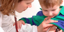
Doctor Banner

An intraventricular hemorrhage can happen with little warning. The result of a brain bleed is often damage that impairs the brain’s ability to control cognitive and motor functions. There are four stages of hemorrhages, all of which can create significant dangers to newborns and infants, requiring delicate surgery.
Hemorrhaging inside the brain, clinically referred to as intraventricular hemorrhage, or IVH, can damage or kill areas of the brain crucial to development and motor function. When this happens, resulting impairment — as well as the severity of impairment — is dependent upon the location and degree of damage. The hemorrhage can be arterial or venous.
Hemorrhaging is bleeding. When bleeding is isolated in a particular organ or tissue, localized swelling, known as a hematoma, can occur. A hematoma can damage and kill surrounding tissue by compressing the tissue or reducing its blood supply. Clotting mechanisms or swelling that block blood supply will eventually stop a hematoma.
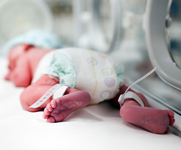
Intraventricular hemorrhage is a significant risk factor for Cerebral Palsy. It is most common in premature babies, especially those who have experienced respiratory distress syndrome, collapsed lung, or high blood pressure. Intraventricular hemorrhage occurs most frequently in the first 48 hours after birth and with diminished likelihood as the infant ages. Intraventricular hemorrhage is categorized into four grades (Grades I through IV) of increasing severity.
The swelling and obstruction, in turn, can lead to a condition called hydrocephalus — increased fluid in the brain — which causes dangerous pressure and may require surgical procedures to relieve.
Changes in blood pressure can also cause delicate infant blood vessels to rupture. Infants placed on ventilators are of particular concern; although great care is taken to prevent rupture, it is possible the child can breathe out of sync with the respirator and cause increased pressure in the lungs and brain.
Risk factors for intraventricular hemorrhage include:
Most common signs of intracranial hemorrhage include:
It is recommended that infants born prematurely receive a routine cranial ultrasound between 7 and 14 days of life, with a second administered at the baby’s original due date. Additional ultrasounds are ordered if a premature baby has new signs or symptoms, especially if his or her health worsens suddenly. It is estimated that an ultrasound will detect problems requiring additional follow-up in 25% of babies born before 30 weeks gestation.
The five forms of intraventricular hemorrhage are:
Intraventricular hemorrhage and hyperbilirubinemia, an elevated level of bilirubin in the blood, pose a strong risk for the development of Cerebral Palsy. This type of hemorrhage is usually caused by an intraparenchymal bleed in the corpus callosum, the bridge of nervous tissue that connects the two cerebral hemispheres allowing communication between them. Upon examination, blood is usually found in the lateral ventricles. Intraventricular hemorrhage is more commonly associated with premature babies who have been exposed to physical stress from respiratory distress syndrome, high blood pressure, or abnormal presence of air or gas in the lungs that leads to a collapsed lung. This condition may occur, on rare occasions, in full-term babies but is rarely present at birth. Head injury, shaken baby syndrome, or fetal stroke are common causes of intraventricular hemorrhage. Risk factors that increase the likelihood of fetal stroke include placental blood clots, malformed or weak blood vessels in the brain, maternal high blood pressure (hypertension), maternal infection, pelvic inflammatory disease, or blood-clotting abnormalities.
Epidural hematoma is the result of ruptured arteries and superficial venous sinuses. It is most often associated with sudden head trauma or accident. While the brain is unharmed, an underlying fracture exists in 60 to 90% of epidural hematomas. Surgery is often required to remove the hematoma and relieve pressure. In children, the classic symptom of brief and gradual loss of consciousness is rare. In children over 6 years of age, it is believed that an injury to the side of the head, such as that sustained in a bicycle fall, is the most common event that can result in epidural hematoma.
Subdural hematoma is generally a more serious condition than an epidural hematoma and is often a component in more severe head injuries. Subdural hematoma is caused by direct trauma, severe acceleration-deceleration trauma or a shaking injury. In children, subdural hematomas may result from birth trauma, child abuse, or shaken baby syndrome. Clinical signs of subdural hematoma in infants include irritability, lethargy, vomiting and bulging soft spot. In cases of abuse, the child will often incur an immediate loss of consciousness, headache, personality change, neck stiffness, seizures, vomiting, low-grade fever, and papillary dilation. Typically, some recovery may occur after head trauma and loss of consciousness, but the child is unlikely to return to his or her normal state. The mortality rate for those with subdural hematoma is significant due to the high incidence of associated irreversible brain damage.
Subarachnoid hematoma is venous (relating to the veins). It is the most common type of intraventricular hemorrhage following birth trauma. Intraventricular hemorrhages resulting from birth trauma are most commonly subarachnoid (bleeding in the head between two membranes surrounding the brain). Subarachnoid hematomas result in the classic posterior interhemispheric region similarly found in cases of shaken baby syndrome but will also hemorrhage in the subarachnoid space, as well. A baby with subarachnoid hematoma will likely develop seizures within 48 hours of birth. The baby may appear to have a stiff neck and be lethargic. Medical practitioners will likely use a lumbar puncture to rule out meningitis before diagnosing this type of hematoma. A non-contrast-enhanced CT scan is often used to detect up to 90% of all subarachnoid bleeds within the first 24 hours of injury.
Intracerebral hemorrhage (ICH) is the bursting of blood vessels between the brain and skull, which often occurs when the brain is unable to absorb the force of impact from a head injury. Neurological damage may occur if additional bleeding occurs and contains cerebrospinal fluid. The two most common areas of the brain to experience this type of hemorrhage are the anterior portion of the temporal lobe and the posterior portion of the frontal lobe. Although the head injury can appear minor, the hematoma can be life-threatening and often requires immediate surgical procedures to relieve pressure caused by compressed brain tissue. This type of hemorrhage is not common in children.
Because the terminology used is so specific, yet remarkably similar, terms such as brain defect, brain malformation and brain lesion can seem confusing. It is helpful to know the difference between the terms when attempting to understand the cause of cerebral palsy.
Brain development begins shortly after conception. A relatively small number of cells divide and multiply into billions of cells. A small strip of tissue rolls into a neural tube. One end develops into the brain, the other into the spinal cord. Throughout, different types of cells form, group, and migrate to form various regions of the brain. The brain is considered fully developed two to five years after birth.
Brain defects are irregularities in the brain structure that typically cause impairment. Defects can occur from malformation, injury, illness or disease. The degree of impairment often is linked to the severity of damage. A brain sometimes compensates for defects, in essence, by “rewiring” to bypass or compensate for damaged areas. For this reason, beginning treatment as early as possible is typically recommended.
Brain malformations are defects that occur through abnormal development of the brain. Although defects can occur anytime during fetal development, in the first 20 weeks the infant is most vulnerable; any malformation that occurs while the neural tube is forming can have permanent consequences. Brain malformations may result in undeveloped areas, abnormal growth, malformation, or improper brain division into hemispheres and lobes.
Brain lesions are defects that occur from injury or disease. The cause of brain lesions during fetal development include bleeding in the brain, infections, toxins, asphyxia, and many others. Lesions typically result from an incident or event that causes brain tissue to die. Holes, which often fill with liquid, are left behind to form cysts.
Cerebral Palsy is caused by brain injury or brain malformation that occurs before, during, or immediately after birth while the infant’s brain is under development. But how a brain injury affects a child’s motor functioning and intellectual abilities is highly dependent on the nature of a brain injury, where the damage occurs, and how severe it is.
Cause
Damage to the white matter tissue in the brain
Periventricular Leukomalacia
Brain hemorrhage
Intraventricular Hemorrhage
Lack of oxygen to the brain, asphyxia, or intrapartum asphyxia
Hypoxic-Ischemic Encephalopathy
Brain malformation or abnormal brain development.
Cerebral Dysgenesis

Cerebral Palsy risk factors are events, substances or circumstances that increase the chances of a child developing Cerebral Palsy. They can be avoidable, or unavoidable. A risk factor does not ensure a child will develop Cerebral Palsy; it means chances are higher than if that risk factor was not present. Likewise, the absence of risk factors does not ensure that a child will not develop Cerebral Palsy. Have you been exposed to the following risk factors?
Cerebral Palsy Risk Factors
Risk factors vs. risk factor causal pathways
A risk factor does not ensure a child will develop Cerebral Palsy; it means chances are higher than if that risk factor was not present. Likewise, the absence of risk factors does not ensure that a child will not develop Cerebral Palsy.
Risk Factors and Risk Factor Causal Pathways
Any exposure to risk factors prior to conception and during pregnancy should be immediately discussed with a doctor in order to treat and minimize risk. The Cerebral Palsy Risk Factor Checklist helps parents determine if they may have been exposed to risk factors for Cerebral Palsy.
The Cerebral Palsy Risk Factor Checklist